Intracellular NK Cell Activation Flow Cytometry Panel R&D Systems

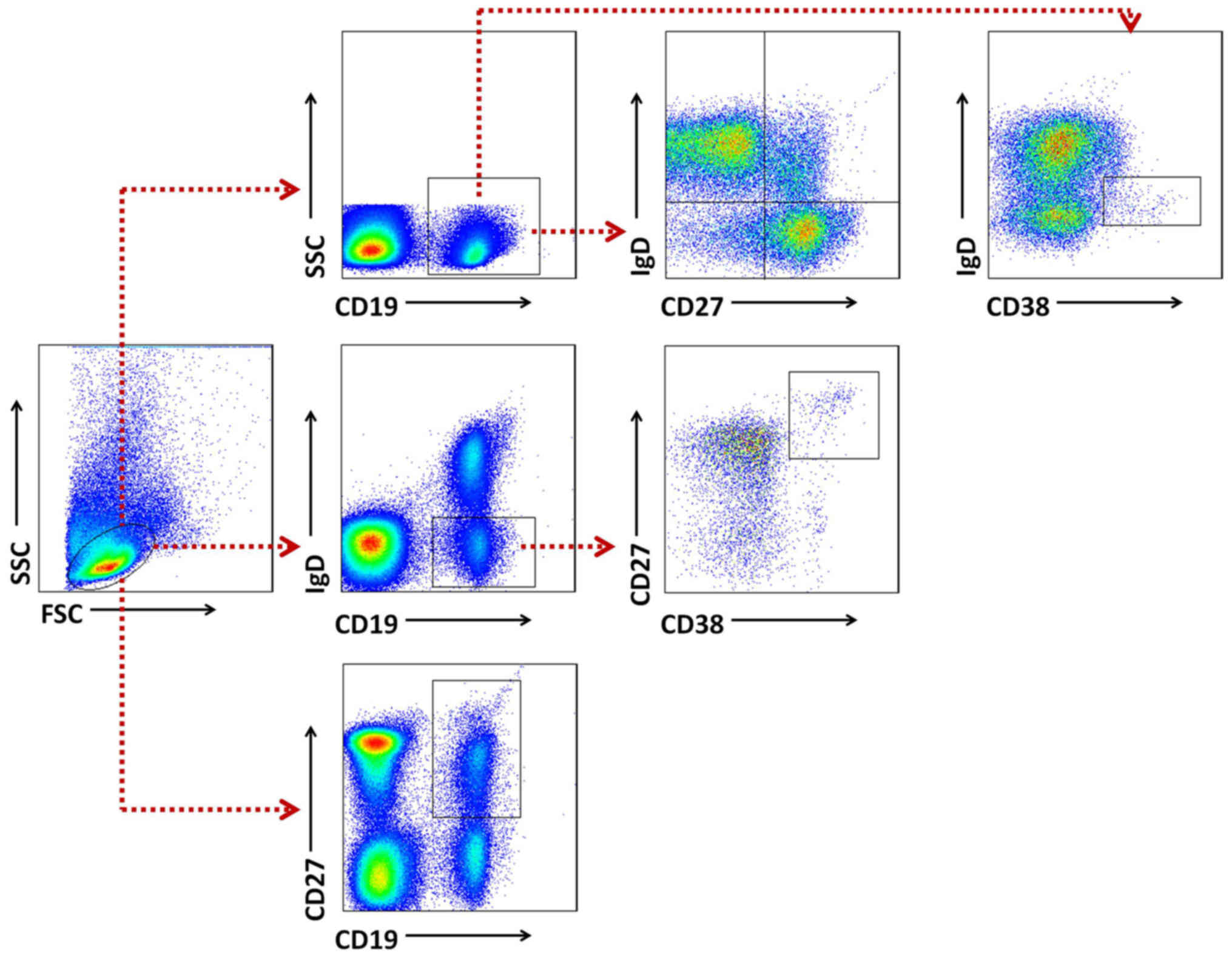
identification of B cells in peripheral blood. (A) Flow cytometry plots showing a gating strategy used to identify CD19 + B cells and CD19 + CD27 hi CD38 hi ASCs.Data are from PBMCs from a representative healthy control donor (HCD). (B) A gating strategy of mass cytometry data to identify live CD19 + B cells.Antibodies to CD3 and CD14 were conjugated to the same metal and therefore created a.
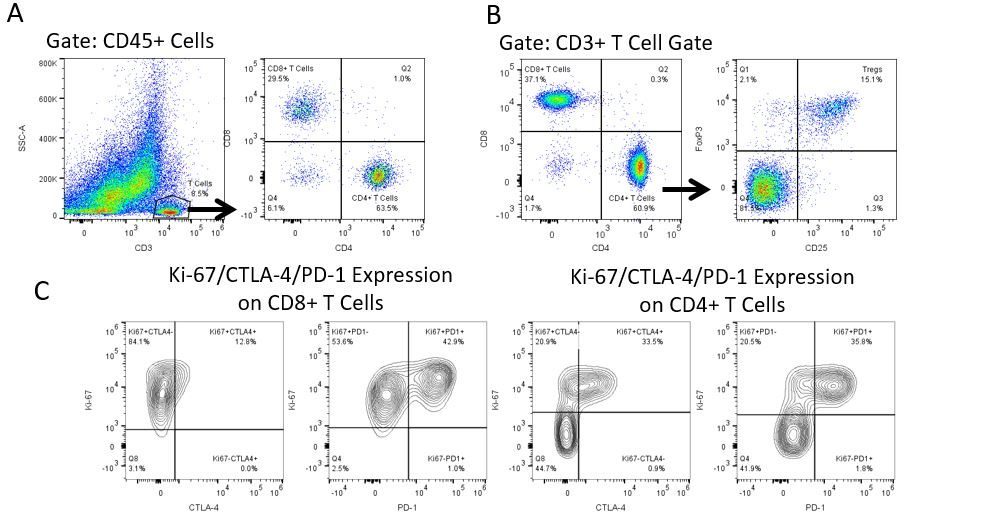
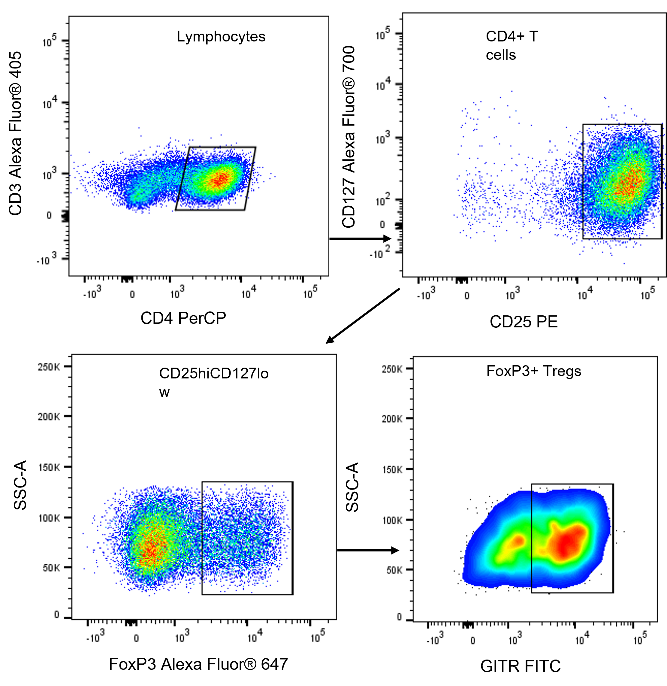
Representative flow cytometry data. (A) Gating scheme for CD4 Tcell... Download Scientific

Viable single cells from mouse spleens are easily obtained using the gentleMACS™ Technology. For downstream flow cytometric analysis of B cells, a validated multicolor flow cytometry panel was designed using REAfinity™ Antibodies and Viobility™ Fixable Dyes. In addition, an optimized gating strategy is provided for the analysis of innate.
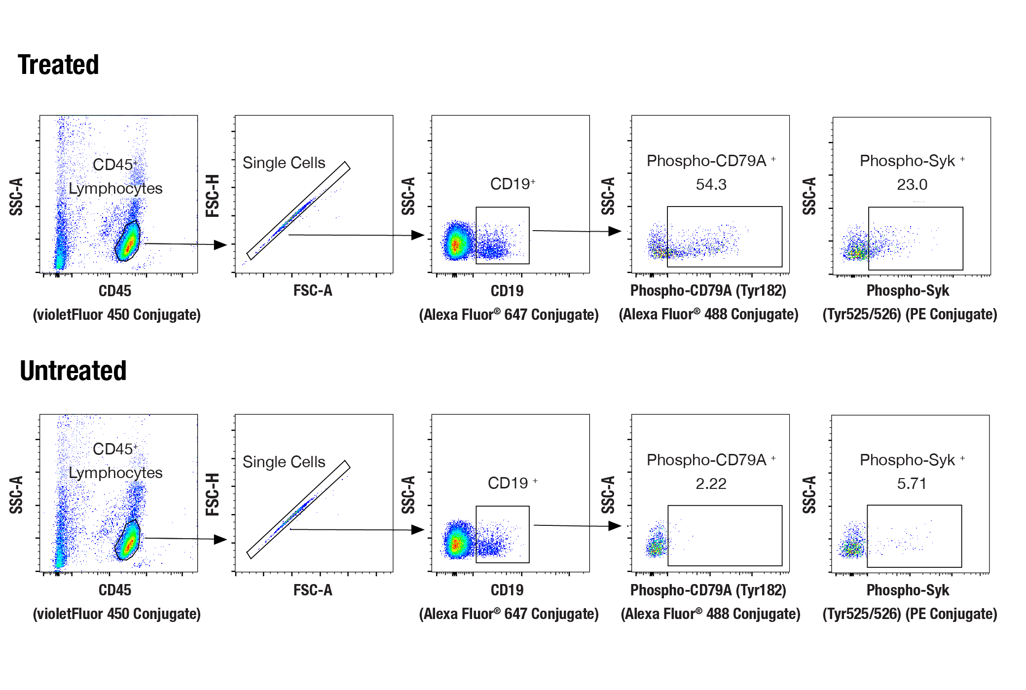
Human B Cell Signaling Flow Cytometry Panel Cell Signaling Technology
To support the use of multicolor flow cytometry for the study of B cells, BD offers a wide portfolio of reagents for B cell phenotyping. They are available in multiple formats, to provide maximum flexibility in panel design.. for panels to do more detailed analysis. The key marker for B cell panels is the lineage marker CD19, which is.
Representative flow cytometry dot plot and gating hierarchy used to... Download Scientific Diagram

The advantage of flow cytometry is that it allows absolute and relative counting of B cells, and their phenotypic and functional evaluation at a single-cell level, while allowing the analysis of a large number of cells. Although versatile and broad in its utility, clinical flow cytometry has both theoretical and practical limitations.
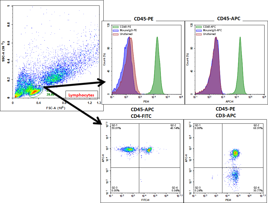
PBMC Immunophenotyping by Flow Cytometry AAT Bioquest
In most cases (e.g. B-cell and T-cell panels), automated analysis provided matched or lowered variability compared to manual analysis (e.g. plasmablasts, transitional B-cells, CD8 central memory.
B Cell Immunophenotyping Cytokine Transcription Detection
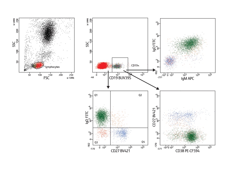
B cell subsets can be difficult to identify due to variability and low level expression of markers and the rarity of some populations. This guide to immunophenotyping of human B cells takes you through some of the common markers and gating strategies used to identify B cells by flow cytometry, with examples of data acquired on the ZE5™ Cell.
Flow Cytometry Gating Strategy Of B Cell Monocyte Myeloid Cell The Best Porn Website

Llinàs, L. et al. Expression profiles of novel cell surface molecules on B-cell subsets and plasma cells as analyzed by flow cytometry. Immunol. Lett. 134 , 113-121 (2011).
B cell development in nanomice a, Flow cytometry analysis of bone... Download Scientific Diagram

This 28-color flow cytometry panel focuses on B cells, dendritic cells and monocytes and was optimized for cryopreserved peripheral blood mononuclear cells (PBMC).. These subsets were combined in the same flow cytometry panel based on shared phenotypic markers such as the high expression of MHC class II molecules, co-stimulatory molecules.
Flow Cytometry Services
For this purpose, we have developed specialized B cell reagent panels for multiparameter flow cytometry, and combine their use with advanced bioinformatics strategies that together will likely be advantageous for improving the characterization, prognosis, and for possibly improving treatment regimens of chronic inflammatory diseases such as lupus.
Increased B cell activating factor is associated with B cell class switching in patients with
Transfer each sample from the 96 well plate to a 5 mL flow cytometry tube. Pass through a cell strainer cap to ensure single cell suspension and wash out with 50-100 μL of staining buffer.. or to enrich B cells from PBMCs prior to the flow cytometric staining, e.g., via magnetic activated cell sorting (MACS). 12, 13. Problem 5. We.
B cell ALL flow cytometry
TLR stimulation of mouse B cells induces class switch DNA recombination (CSR) to isotypes specified by cytokines, and also induces formation of IgM(+) as well as c. Analysis by Flow Cytometry of B-Cell Activation and Antibody Responses Induced by Toll-Like Receptors Methods Mol Biol. 2016:1390:229-48. doi: 10.1007/978-1-4939-3335-8_15..
Flow Cytometry Panel Products R&D Systems
In this review, we summarize the established techniques and discuss new and emerging technologies for probing the B cell response in vitro and in vivo by taking advantage of the specificity of B cell receptor (BCR)-associated and secreted antibodies. These include ELISPOT, flow cytometry, mass cytometry, and fluorescence microscopy to identify.
Human CD19 B Cell Flow Cytometry Panel FMCP003 R&D Systems

Some B cell subsets are defined by their function. First, the activation of B cells can be assessed by flow cytometry. Several intracellular and surface markers are associated with activation. In the panel used throughout this paper, downregulation of CD24 and CD38 can mark activated cells.
Expression of Lselectin (CD62L) by normal Bcells and BCLL cells.... Download Scientific Diagram
Flow cytometry using fluorescent hemagglutinin and B cell marker specific Abs enables high throughput qualitative and quantitative detection of individual B cells. By using a graded amount of antigen and gating on GC B cells we define the AC 50 the amount of antigen required to stain 50% of hemagglutinin specific B cells. This number is in.
mRNA expression levels of Bcell receptor components in normal and... Download Table

This prompts to perform immunophenotypic investigations that will reveal, in most cases, the presence of monotypic B cells. Multiparameter flow cytometry (MFC) thus plays a critical role in the diagnosis and characterization of B-cell LPDs with PB and/or bone marrow (BM) involvement.. the orientation panel can usefully include CD5, CD20,.
Immunophenotypic analysis by flow cytometry revealed a common Bcell... Download Scientific

Identification and characterization of B-cells by flow cytometry. (A) Identification of B-cells (blue) using CD19 (left) and CD20 (right). Utilization of an alternative B-cell marker such as CD40 (panel F) allows clear discrimination of the neoplastic cells from CD5+ T cells. (Fig. 4 courtesy Dr. Sindhu Cherian, University of Washington.
.

